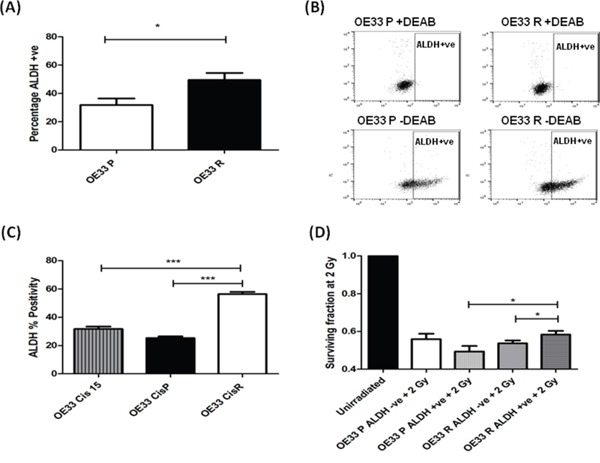
Figure 3. Radioresistant and cisplatin-resistant EAC cells have increased ALDH enzymatic activity, and is associated with a radioresistant phenotype.

A. OE33 P and OE33 R cells were stained for ALDH and fluorescence was assessed using a MoFlow cell sorter. As a negative control, cells were treated with the specific ALDH inhibitor DEAB. OE33 R cells demonstrate significantly higher levels of ALDH+ve cells, when compared to OE33 P cells. Data are presented as the mean ± SEM from 7 independent experiments. Statistical analysis was performed using an unpaired two-tailed Student's t-test, *p < 0.05 B. Representative flow images of ALDH staining in OE33 P and OE33 R cells. DEAB served as a negative control. C. ALDH activity was assessed in an isogenic model of EAC cisplatin-resistance. Cisplatin-resistant cells OE33 CisR demonstrated significantly higher levels of ALDH+ve cells, when compared to cisplatin-sensitive OE33 CisP and OE33 Cis15 cells. Data are presented as the mean ± SEM from 3 independent experiments. Statistical analysis was performed using an unpaired two-tailed Student's t-test, ***p < 0.001 D. ALDH-ve and ALDH +ve populations from OE33 P and OE33 R cells were sorted using a MoFlow cell sorter and radiosensitivity to 2 Gy X-ray radiation was assessed using clonogenic assay. OE33 R ALDH+ve populations demonstrate significantly enhanced survival to radiation at 2 Gy, when compared to OE33 R ALDH-ve cells and OE33 P ALDH+ve cells. Data are presented as the mean ± SEM from 7 independent experiments. Statistical analysis was performed using a paired and unpaired two-tailed Student's t-test, respectively, *p < 0.05.
