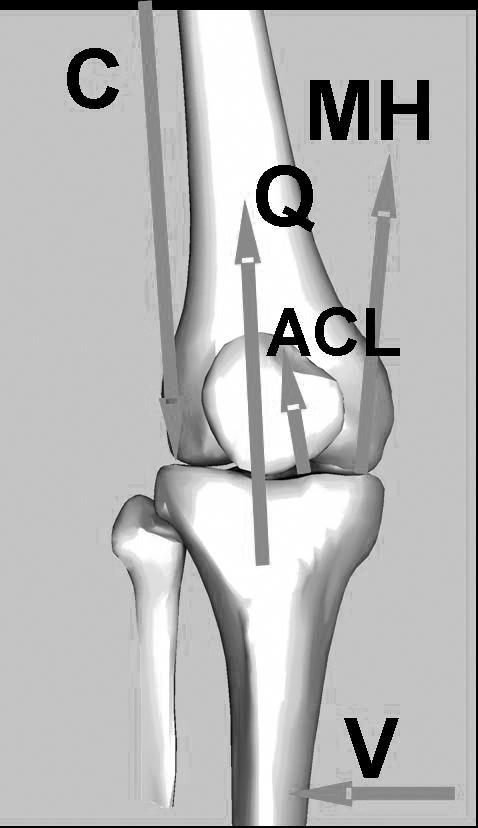
Figure 1. Free-body diagram of forces acting on the tibia, showing the frontal-plane equilibrium between external valgus load (V), articular contact force (C), quadriceps force (Q), medial hamstrings (MH), and anterior cruciate ligament (ACL) force.

Under external valgus loading, contact shifts to the lateral compartment. The moment balance with respect to the contact point shows that Q and MH both help the ACL (and the medial collateral ligament, not shown) stabilize the joint against valgus loading. Under a given valgus load, any reduction in these muscle forces increases ligament loading
