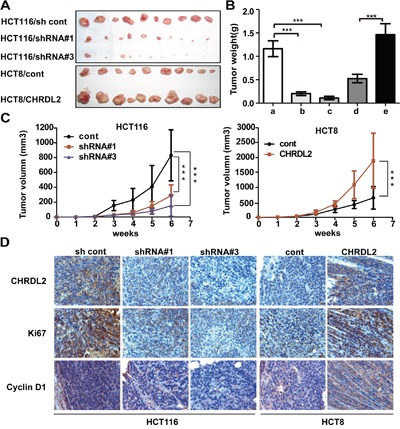
Figure 5. The effect of CHRDL2 on tumor growth of subcutaneous xenograft of CRC cells in nude mice.

A. Lentivirus-transfected CRC cells (HCT116/sh control, HCT116/shRNA#1, HCT116/shRNA#3, HCT8/cont, HCT8/CHRDL2) were used for in vivo xenograft tumor growth assay, with 2×10⁶ cells being subcutaneously injected into a flank of nude mice. B. The histograms show the weight of xenograft tumors (**P<0.01, ***P<0.001). C. Cure graph showing the volume of xenograft tumors forevery week during6 weeks (**P<0.01, ***P<0.001). D. IHC staining of CHRDL2, Ki67, and cyclin D1 expression in subcutaneous xenograft tumor. The scale bar represents 100 μm.
