
Figure 3. Immunohistochemical and cytogenetic features of B-cell lymphomas involving WR.
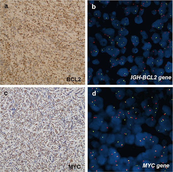
(A, B), case 8, PDLBCL. (A) BCL2 was positively expressed in neoplastic cells. b, FISH analysis did not detect a positive fusion signal in BCL2 of the samples. LSI IGH/BCL2 dual-color, dual-fusion probe signal pattern expected for t(14;18)(q32;q21). SpectrumOrange-labeled BCL2 probe and SpectrumGreen-labeled IGH probe correspond to 18q21 and 14q32, respectively. (C, D), case 3, BL. (C) MYC was positive expression. (D) Presence of LSIMYC Dual Color, BreakApart Rearrangement Probe signal pattern with abnormal nucleus showing a one orange, one green and one orange/green fusion signal pattern. Normal probe hybridizes to the band region 8q24. SpectrumOrange probe begins upstream of the 5′ end of MYC and SpectrumGreen probe starts downstream of 3′ of the MYC gene.