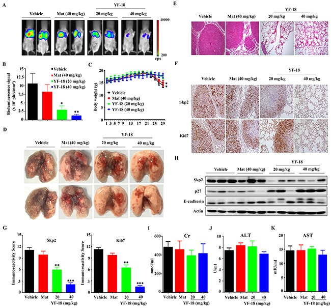
Figure 6. In vivo anti-lung cancer efficacy of YF-18.

A. A549-Luciferase cells were intravenously injected into SCID mice, and 4 days later the mice were randomized to receive vehicle, matrine, and YF-18 treatment (n = 6 for each group). The mice were detected by IVIS Spectrum. B. The relative luciferase intensity in the mice. C. The body weight of mice was monitored every two days. D. Representative images of dissected lung tissue from each group. E. Hematoxylin and eosin (HE) staining of lung tissue sections of mice from each group. F. Immunohistochemistry (IHC) using anti-Skp2 and Ki67 antibodies. G. Statistic analysis of IHC staining. H. Western blot assays using lysates of isolated tumors and indicated antibodies. I-K. The serum Cr (I), ALT (J) and AST (K) levels of mice from each group were detected. Data is represented as mean ± SD.
