
Figure 2. Establishment and characterization of LOXL4 knockdown MDA-MB-231 cells.
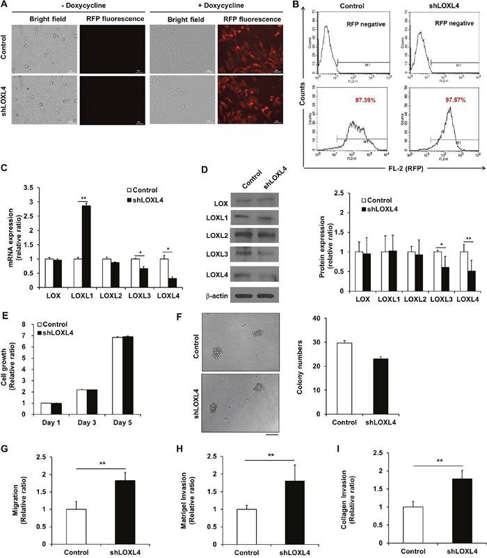
A. Fluorescence images of doxycycline-inducible red fluorescent protein expression in MDA-MB-231 cells transduced with lentivirus encoding both RFP and either the non-silencing control vector or shLOXL4. Scale bar: 50 μm. B. Flow cytometric analysis of the percentage of RFP-positive control or LOXL4-knockdown MDA-MB-231 cells. C. Quantitative real-time RT-PCR of LOX and LOXL1-4. D. Western blotting analysis of LOX family member protein expression. E. MTT assay for analysis of cell proliferation. F. Single cell colony formation assay. Scale bar: 100 μm. G. Trans-well migration assay for the analysis of cell migratory capacity. H and I. Trans-well invasion assays for the analysis of cell invasion capacity. All experiments were performed at least in triplicate; means ± standard deviation of all experiments are shown. *P < 0.05. **P < 0.001. Scale bar: 50 μm.