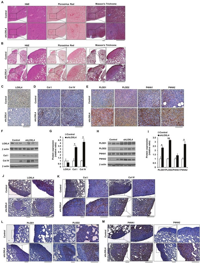
Figure 4. LOXL4 knockdown increases collagen synthesis and deposition.

A. Images of H&E-, Picrosirius red-, and Masson's trichrome-stained sections of primary tumors resulting from injection of control and LOXL4-knockdown cells. Scale bar: 400 μm. B. Images of H&E-, Picrosirius red-, and Masson's trichrome-stained lung sections after injection of control and LOXL4-knockdown cells. Scale bar: 400 μm. C and D. Representative immunohistological (IHC) images of LOXL4, collagen I, and collagen IV staining in primary tumors. Scale bar: 100 μm. E. Representative IHC images of PLOD1-2 and P4HA1-2 staining in primary tumors. Scale bar: 100 μm. F. Representative images of Western blots for LOXL4, type I procollagen (collagen I), and collagen IV. G. Densitometric quantification of LOXL4, type I procollagen (collagen I), and collagen IV expression in tumors. H. Western blotting analysis of PLOD1-2 and P4HA1-2 expression in primary tumors. I. Densitometric quantification of PLOD1-2 and P4HA1-2 expression in tumors. J and K. Representative IHC images of LOXL4, collagen I, and collagen IV staining in the lungs. Scale bar: 100 μm. L and M. Representative IHC images of PLOD1-2 and P4HA1-2 staining in the lungs. Scale bar: 200 μm. *P < 0.05. **P < 0.001.
