
Figure 2. Pax-5 is expressed in clinical breast cancer tissues.
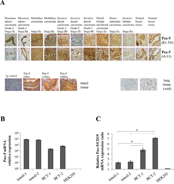
A. Immunohistochemistry was performed on FFPE breast tissue microarrays representing a panel of 306 core samples from healthy and varying mammary cancer types. Tissues slides were probed with anti-Pax-5 antibodies targeting the exon 5/6 (Ex 5/6) and N-terminal (A-11) regions respectively and subsequently revealed with HRP-conjugated host antibodies and hematoxylin counterstaining. Images were taken at a 400X magnification and are representative of duplicate samples from tissues cores of 1.5 um in diameter. Control samples include mammary tissues probed with an isotype-matched irrelevant primary antibody (Ig control); tonsil tissues (Pax-5-bearing B cell) used as a positive control; and, cancerous lung tissue as a negative control (-ctrl). Relative mRNA expression levels of Pax-5 B. and Pax-5/CD19 ratios C. were determined by RT-qPCR in clinical FFPE tonsil samples (tonsil-1 and -2) and breast cancer tissues (BCT-1 and BCT-2). HEK293 cells were used as a negative control. The presented data is the calculated mean of three independent samples where statistical analysis by t-test indicates significant differences in respect to control cells (* p<0.01).