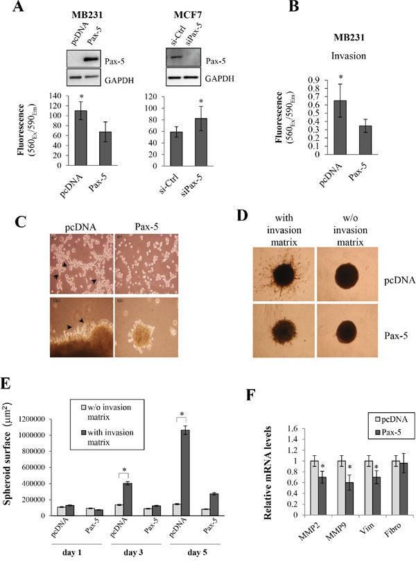
Figure 5. Pax-5 suppresses breast cancer cell malignant features.

A. Cell migration was monitored in MB231cells (left panel/transfected with pcDNA or Pax-5) and MCF7 cells (right panel/transfected with a Pax-5 silencing siRNA (siPax-5) or a control scrambled siRNA (si-Ctrl)). Pax-5 and GAPDH protein expression levels were also validated by Western blot (top panels) for each respective breast cancer models. B. MB231 cells transfected with either Pax-5 or pcDNA were monitored for cell invasion processes and C. anchorage-independent growth. Images were taken at a 40X (top panels) and 100X (bottom panels) magnifications where black arrows point to elongated mesenchymal-like cells. D. Stably transfected BT549 cells with Pax-5 were evaluated for 3D spheroid formation assays with or without (w/o) the presence of invasion matrix and E. quantified using the ImageJ software to assess the total surface invaded by migrating cells. F. Transfected MB231 cells were also monitored for matrix metallo-proteinases (MMP-2 and MMP-9), vimentin (Vim) and fibronectin (Fibro) using RT-qPCR. The presented data is the calculated mean of three independent experiments where statistical analysis was determined by t-test in respect to control cells (* p<0.01).
