
Figure 4. Sub-cultured spheroids.
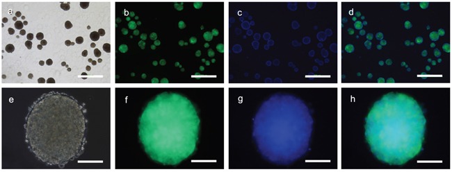
Cells derived from a single primary spheroid developed directly into secondary spheroids without a monolayer in the culture dishes. a. Multiple spheroids were observed three days after subculture which b. expressed GFP and c. consisted of a three dimensional cell masses as seen via staining of the cell nuclei with Hoechst dye. e. The individual spheroids consisted of small cells that f. were GFP positive with g, h. a dense central mass. Panels a and e – phase contrast microscopy. Panels b and f – GFP fluorescence microscopy. Panels c and g – Hoechst fluorescence staining. Panels d and h – merged images. Bars panels a to d = 500μm; panels e to h 100μm.