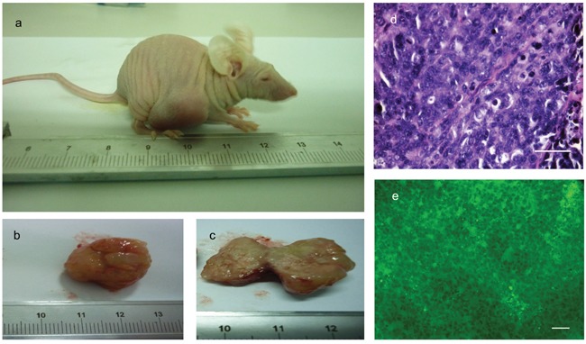
Figure 7. Cells isolated from PIWIL2 expressing spheroids form tumors in athymic mice.

a. Five weeks after subcutaneous injection of cells derived from spheroids tumors were clearly visible at the injection sites. b. Excised tumors consisted of a relatively undifferentiated mass of parenchyma which in cross section c. showed the presence of grey to pink neoplastic tissue with local hemorrhagic necrosis. d. Hematoxylin and eosin staining of thin sections revealed a mass of rather uniform undifferentiated cells with nuclear pleomorphisem, enlarged nuclei, and high nuclear to cytoplasmic ratio. e. These malignant cells were positive for GFP expression as seen by fluorescence microscopy indicating that the tumors originated form the human foreskin fibroblasts originally transfected with PIWIL2 plus GFP that were the source of the secondary spheroids used to obtain the cells that were injected into the mice. There is no evidence of teratoma formation. Bar d = 50μm; e = 100μm.
