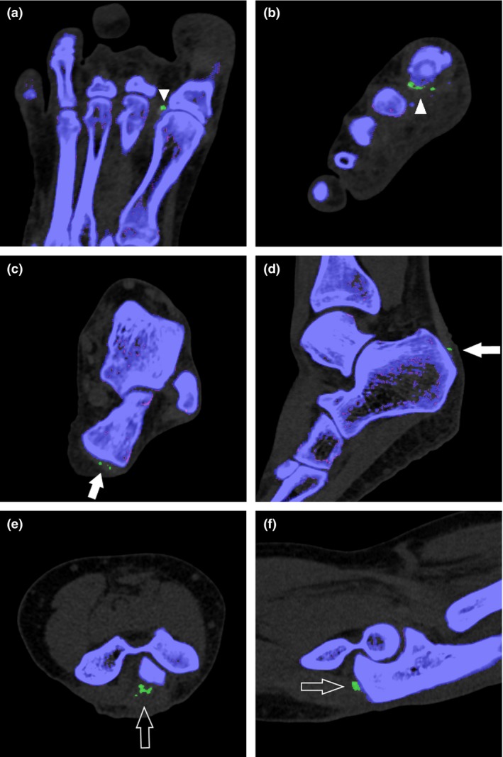
Figure 3.

Post‐processed images in the axial (a) and coronal (b) planes demonstrate typical monosodium urate (MSU) deposits along the lateral aspect of the first metatarsophalangeal joint (arrowheads). Axial (c) and sagittal (d) images show MSU deposits along the distal end of the Achilles tendon (arrows). Axial (e) and sagittal (f) images depict MSU deposits along the distal triceps tendon (open arrows).
