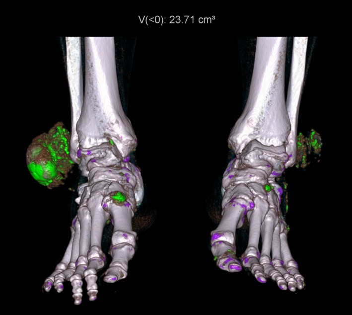
Figure 7.

Three‐dimensional rendered image depicting large tophi over the lateral malleoli of both ankles as well as smaller deposits scattered around both ankles and feet. Automated quantification of urate volume is displayed at the top of the image.
