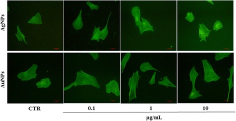
Fig. 3.

Cytoskeletal reorganization in human fibroblasts treated with NPs. Cells exposed to AuNPs and AgNPs at 0.1, 1, or 10 μg/mL for 24 h were stained with phalloidin to analyse F-actin distribution (green staining). Photomicrographs revealed alterations in cell polarity and stress fibres in NP-treated cells. CTR: non-treated cells. Magnification, 200×
