
FIGURE 7.
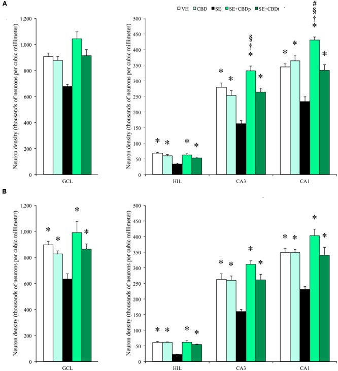
Cannabidiol effects on hippocampal neuronal survival, 1 day after SE. Neuronal density, estimated in contralateral (A) and ipsilateral (B) hippocampal sections from VH (white bars), CBD (light green bars), SE (black bars), SE+CBDp (medium green bars), and SE+CBDt (dark green bars) groups. All groups had higher neuron density than SE group in the hilus, CA3, and CA1 subfields of contralateral (A) and ipsilateral (B) hippocampus (ANOVA followed by Sidak post hoc test). In the contralateral hippocampus, SE+CBDp group had higher neuron density than CBD and SE+CBDt in CA3 and CA1. SE+CBDp had also higher neuron density than VH group in the contralateral CA1 subfield. The groups VH, CBD, SE+CBDp, and SE+CBDt had also higher neuron density than SE group in the ipsilateral granule cell layer. GCL, granule cell layer; HIL, hilus. The ∗ indicates differences from SE group, the † indicates difference from SE+CBDt group, the § indicates difference from CBD group, and the # indicates difference from VH group. Data were presented as mean ± standard error.