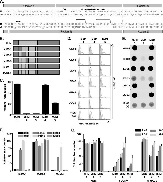
FIG 5.
Region 4 within GP1 plays a critical role in RBS accessibility for α-JUNV neutralizing antibodies. (A) MACV (Carvallo; M) or JUNV (Candid#1; J) GP1 amino acid sequences aligned according to conserved cysteine residues. The three amino acid residues that differ in the Candid#1 strain from the recXJ13 strain of JUNV are underlined. Conserved cysteines are indicated by gaps in region labels, and partners in disulfide bonds for all cysteine residues are indicated by connecting lines. The five regions exchanged to form the various intraGP1 MJM chimeras are indicated by the labeled gray bars above and below the sequences. Circles marking specific residues within the sequences are presented as indicated in Fig. S2A in a study by Mahmutovic et al. (28). White circle indicates JUNV GP1 residue predicted to only contact TfR1, gray circles indicate JUNV GP1 residues only contacted by GD01, and black circles indicate JUNV GP1 residues that are both predicted to contact TfR1 and interact with GD01. (B) Diagram of MJM intraGP1 chimera assembly and nomenclature. (C) Analysis of transduction competence among various MJM intraGP1 chimera-VSV pseudovirions. Pseudovirion preparations were normalized to MJM-VSV for VSV matrix expression by dot blot analysis and applied to Vero cells in duplicate. Transduction is represented as the percentage of GFP-positive cells relative to MJM-VSV, as assessed by flow cytometry. Bars are representative of three independent pseudovirion preps with the indicated SEM. (D) Cell surface expression analysis of MJM intraGP1 chimera-expressing 293T cells. Cells were transfected with GPC-containing plasmids 48 h prior to staining with a 1:50 dilution of each α-JUNV GPC MAb or a 1.5:2 dilution of F106G3. Filled histograms represent cells transfected with empty vector; unfilled histograms represent cells transfected with respective GPC plasmids. (E) Dot blot analysis of nondenatured MJM intraGP1 chimera-VSV-associated GPCs. Pseudoviruses were incubated with lysis buffer prior to direct application to nitrocellulose. Blots were incubated with either a 1:500 dilution of each α-JUNV MAb or a 1:20 dilution of F106G3. (F) Neutralization assays assessing the capacity of each α-JUNV MAb to inhibit MJM intraGP1 chimera-VSV transduction. Pseudovirions were incubated with each MAb at 0.5 μg/ml prior to application to Vero cells. Transduction is represented as the percentage of GFP-positive cells relative to cells incubated with nonspecific MAb, as assessed by flow cytometry. Bars are representative of two or three independent experiments with the indicated SEM. (G) Neutralization assays assessing the capacity of polyclonal antisera (α-JUNV, α-MACV, or NMS) to inhibit the transduction of each MJM intraGP1 chimera-VSV. Pseudovirions were incubated with decreasing concentrations of each antiserum type or PBS prior to application to Vero cells (dilution factors were as indicated in the inset key). Transduction is represented as the percent of GFP-positive cells relative to cells receiving PBS treatment, as assessed by flow cytometry. Bars are representative of two or three independent experiments with the indicated SEM.

