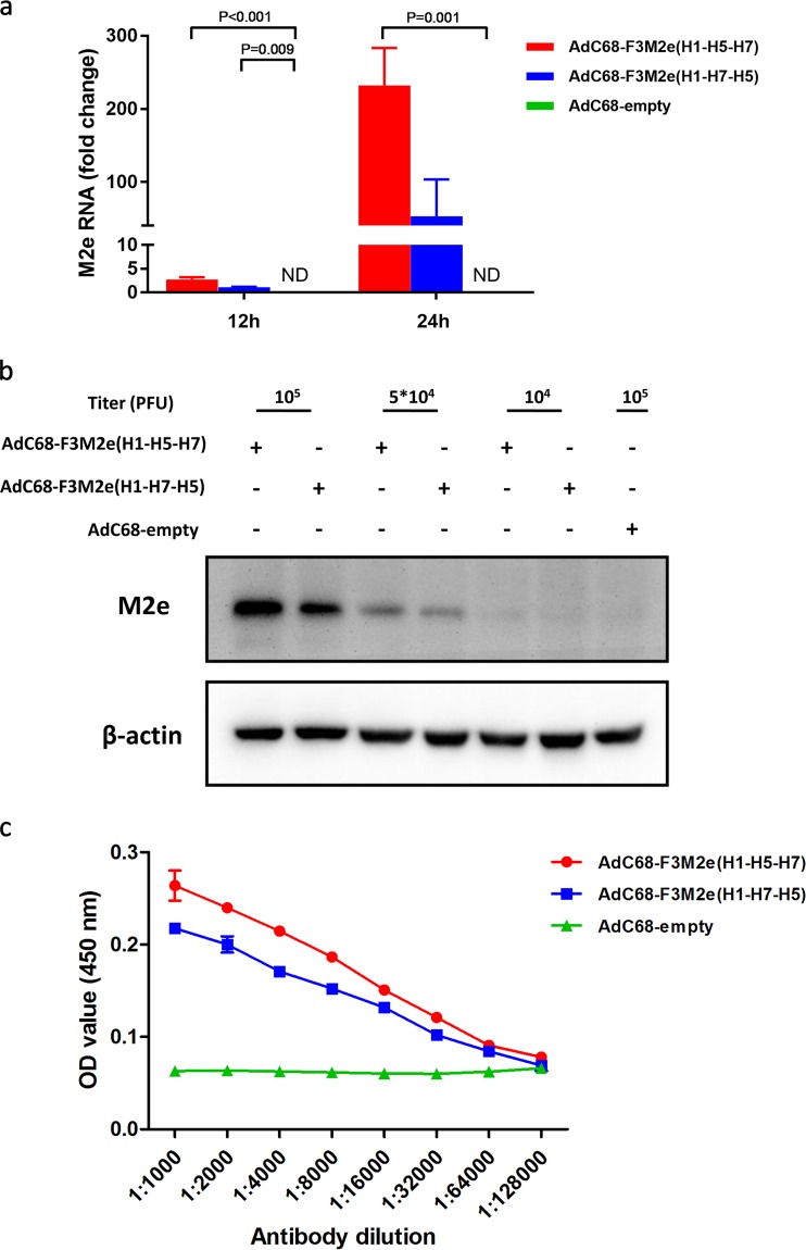
FIG 3.
Expression of M2e incorporated in AdC68 fiber. (a) HEK293 cells were infected with AdC68-F3M2e(H1-H5-H7), AdC68-F3M2e(H1-H7-H5), or AdC68-empty at an MOI of 0.1. Total RNA was extracted and applied for M2e RNA expression analysis by real-time PCR 12 or 24 h later. Values were normalized to β-actin data and are expressed as average fold changes relative to AdC68-F3M2e(H1-H7-H5) (the reference) ± standard deviations (SD). (b) Different doses of AdC68-F3M2e(H1-H5-H7) or AdC68-F3M2e(H1-H7-H5) were transduced into HEK293 cells and analyzed for M2e expression (relative to β-actin) by Western blotting 24 h later. AdC68-empty served as a negative control. (c) Plates were coated with 5 × 10⁴ PFU of the indicated adenoviruses, blocked, and incubated with 2-fold serially diluted (from 1:100 to 1:12,800) 14C2. HRP-conjugated anti-mouse IgG served as secondary antibody. Data are shown as mean absorbance ± standard deviation (SD). All experiments were repeated in triplicate.

