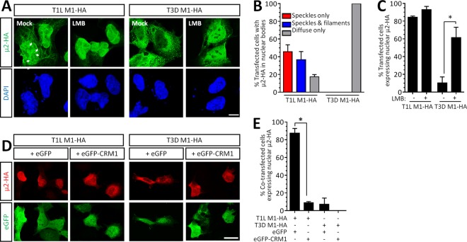
FIG 1.
μ2 undergoes constant CRM1-dependent nuclear shuttling, but the predominant intracellular localization is strain specific. (A) AD-293 cells were transfected with reovirus T1L- or T3D-M1-HA for 20 h and treated with medium alone (mock) or the CRM1 inhibitor LMB for 5 h. Arrows depict μ2 localization to intranuclear bodies. Scale bar, 20 μm. (B) The percentage of cells in panel A displaying the indicated nuclear bodies is presented for four independent experiments (means ± the SEM; n = 6 to 61 cells per condition). (C) The percentage of cells in panel A displaying nuclear μ2 is presented for two to four independent experiments (means ± the SEM; n = 46 to 197 cells per condition). (D) AD-293 cells were cotransfected with reovirus T1L- or T3D-M1-HA and either an eGFP or eGFP-CRM1 plasmid. Scale bar, 10 μm. (E) The percentage of cotransfected cells in panel D displaying nuclear μ2 is presented for two independent experiments (means ± the SEM; n = 18 to 71 cells per condition). *, Significantly different (P < 0.05).

