
FIG 2.
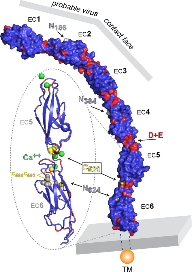
CDHR3 model. Sequences for all 6 EC repeat domains of human CDHR3 were modeled by ACP using I-Tasser procedures similar to those in reference 11. Key residues for Asn-linked glycosylation (gray), acidic residues (red), and Cys locations (yellow) are highlighted. The assignments of Ca2+ coordinates (green) in the EC5-EC6 insert are from alignment with PDB 4ZPM, protocadherin alpha C2 EC1-3.