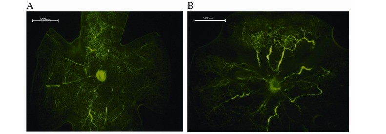
Figure 1.
Retinal angiography with high-molecular-weight fluorescein-dextran. (A) Retina of the control group. No neovascularization was observed in the peripheral perfusion area. (B) Retina of the oxygen-induced retinopathy group. Decreased vascular plexus and vascular tortuosity was visible in the peripheral perfusion area. Original magnification, ×40; scale bars=500 µm.

