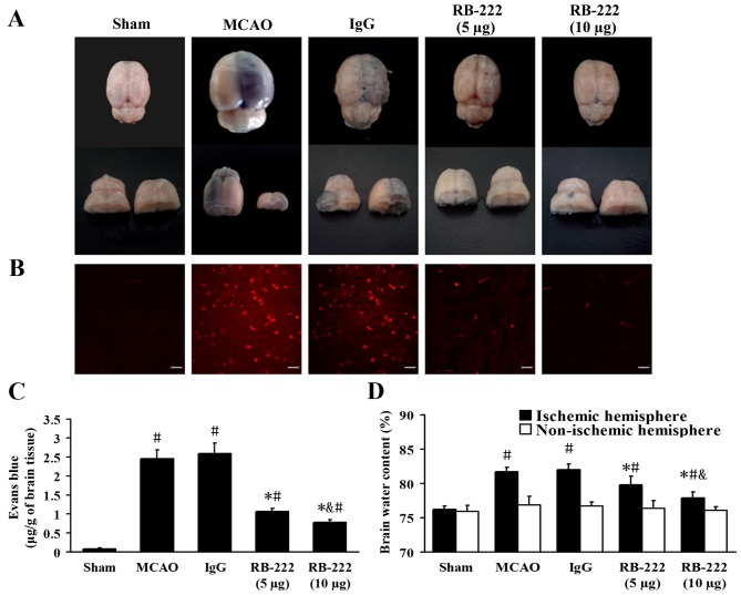
Figure 3.
VEGF inhibition attenuated EB extravasation and brain edema at 24 h after reperfusion onset. (A) Representative images of EB-stained brains at 24 h following MCAO. (B) Confocal microscopy images showing EB dye (red) in each group (scale bar, 20 µm). (C) Quantitative analysis of EB extravasation (µg/g of brain tissue). (D) Brain water content in the ipsilateral hemisphere was significantly decreased following RB-222 treatment. Data presented as the mean ± standard deviation (n=6). #P<0.05 vs. Sham; *P<0.05 vs. MCAO or IgG; &P<0.05 vs. RB-222 (5 µg). VEGF, vascular endothelial growth factor; EB, Evans Blue; MCAO, middle cerebral artery occlusion.

