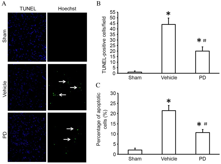
Figure 2.
PD treatment reduces apoptosis in the cortex following middle cerebral artery occlusion. (A) TUNEL staining in the cortex (white arrows indicate the TUNEL-positive cells). Original magnification, ×200 magnification. (B) Quantification of TUNEL-positive cells averaged over 10 microscopic fields per animal. (C) Isolated neuronal apoptosis as evaluated by the Annexin V/PI double stain assay. Data are presented as the mean + standard deviation (n=6 in each group). *P<0.05 vs. sham group; #P<0.05 vs. vehicle group. TUNEL, terminal deoxynucleotidyl transferase dUTP nick end labeling.

