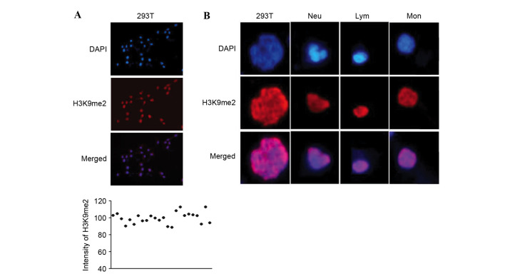
Figure 1.
Immunofluorescence staining of H3K9me2 in 293T cells and three types of WBCs. (A) Homogenous fluorescence staining and intensity of H3K9me2 in 293T cells; DAPI was used to stain the nuclei. The scatter plot indicates that 293T cells demonstrated homogeneous staining. (B) Different nuclear morphology of 293T and three types of WBCs, Neu, Lym and Mon allows distinction of the four cell types. H3K9me2, histone 3 lysine 9 dimethylation; WBCs, white blood cells; Neu, neutrophils; Lym, lymphocytes, Mon, monocytes.

