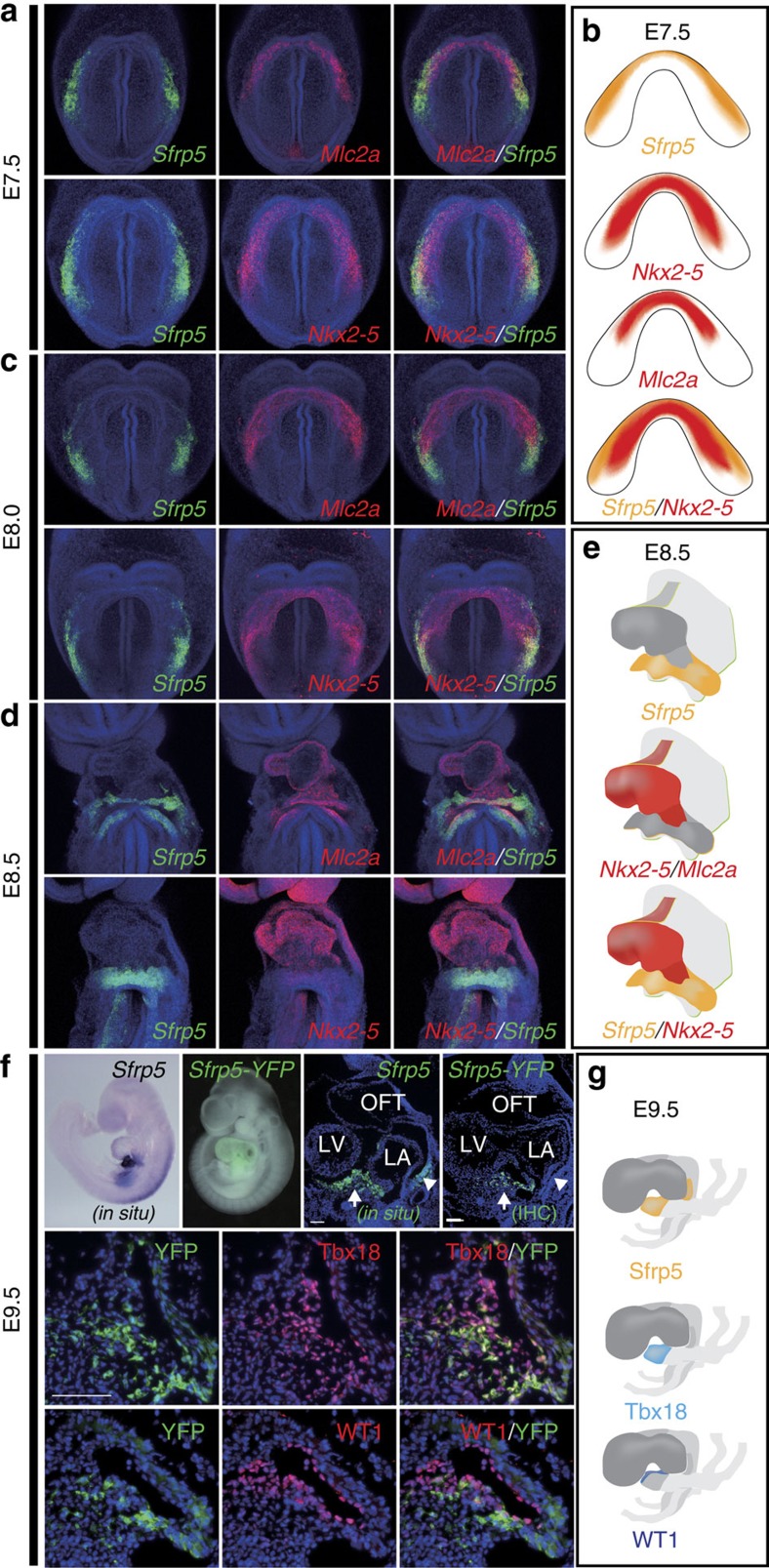
Figure 1. Sfrp5 is expressed in the lateral cardiac crescent and subsequently in the SV.
(a,c,d) Double fluorescent whole-mount ISH of mouse embryos at E7.5 (a), E8.0 (c) and E8.5 (d) for detection of Sfrp5 (green) using Mlc2a or Nkx2-5 (red) as indicated in each panel. (b,e,g) Schematic drawings of gene expression (b,e) and protein distribution (g). (f) Whole-mount ISH (in situ), YFP image merged with bright field image (Sfrp5-YFP), section in situ with probe for Sfrp5 and single or double immunohistochemistry (IHC) of Sfrp5-venusYFP KI embryos using anti-GFP antibodies with anti-Tbx18 or anti-WT1 antibodies at E9.5, as indicated in each panel. Scale bars=Scale bars, 50 μm.

