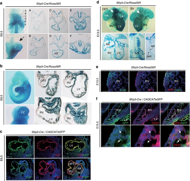
Figure 2. Descendants of Sfrp5-expressing cells contribute to the SV and all chamber myocardium except for the RV.
(a,b,d) Whole-mount and section LacZ staining of Sfrp5-Cre/Rosa26R embryos at E8.5 (a), E9.5 (b) and E13.5 (d). HT: heart tube; dVP: dorsal venous pole; vVP: ventral venous pole; epi: epicardium; peri: pericardium, IVS; interventricular septum. (c,e,f) Double immunohistochemistry of Sfrp5-Cre/CAG-floxed-CAT-eGFP (c,f) or Sfrp5-Cre/Rosa26R (e) embryos at E 9.5 (c), E 13.5 (e) and E15.5 (f), using anti-GFP or anti-β-gal (green) antibodies with anti-TnT, anti-Isl1, anti-WT1 or anti-CD31 antibodies (red). Rectangles in upper panels (f) are magnified in lower panels. Arrows indicate cells co-expressing YFP and CD31 (f). Scale bars=50 μm.

