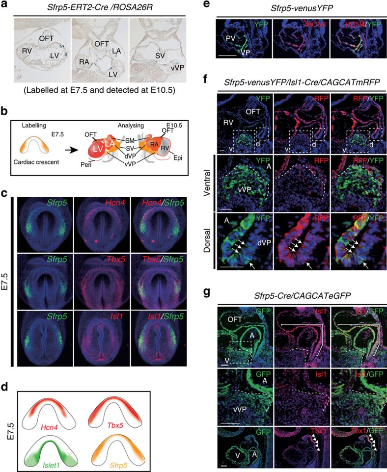
Figure 3. Sfrp5-expressing cells at E7.5 contribute to the SV and all chamber myocardium except for the RV.
(a) Representative photographs of LacZ-stained sections from Sfrp5-ERT2Cre/Rosa-lacZ embryos at E10.5, labelled by tamoxifen at E7.5. (b) Schematic drawing for the expression of Sfrp5 (orange) and the cardiac contribution of its lineages (red) when cells were labelled at E7.5 and analysed at E10.5. (c) Double fluorescent whole-mount ISH of mouse embryos at E7.5 for detection of Sfrp5 (green) and Hcn4, Tbx5 or Isl1 (red), as indicated in each panel. (d) Schematic drawings of gene expression of Sfrp5, Isl1, Hcn4 and Tbx5. (e) Double immunohistochemistry of Sfrp5-venusYFP at E8.5 using anti-GFP (green) and anti-HCN4 (red) antibodies. Co-expression with HCN4 was found in the VP. Weak expression of YFP also co-distributed with HCN4-positive cells in the IFT and part of the primitive ventricle (PV). (f,g) Double immunohistochemistry of Sfrp5-venusYFP (e), Sfrp5-venusYFP/Isl1-Cre/CAG-(floxed)-CAT-mRFP (f) or Sfrp5-Cre/CAG-(floxed)-CAT-eGFP (g) embryos at E9.5 using anti-GFP (green) antibodies and anti-HCN4, anti-RFP, anti-Isl1 or anti-Tbx1 (red) antibodies. Rectangles in upper panels are magnified in lower panels (v: ventral; d: dorsal). Co-expression with Isl1 was detected (arrows in panel (f) and parenthesis in panel (g)). Boundaries for the VP from the atrium are indicated by white dotted lines (f,g). Signal for Tbx1, but not GFP, was observed in the anterior splanchnic mesoderm and anterior OFT (arrowheads in panel (g)). A, atrium.

