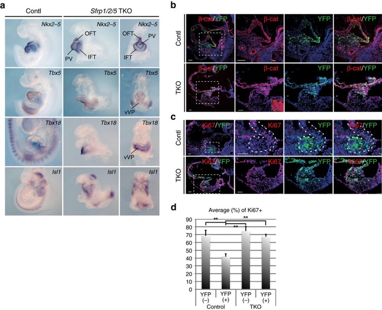
Figure 4. Sfrp1/2/5 TKO embryos exhibit hyperplasia of the IFT accompanied by activated canonical Wnt signalling.
(a) ISH of control and TKO embryos with probes for Nkx2-5, Tbx5, Tbx18 and Isl1 genes. PV: primitive ventricle. (b,c) Double immunohistochemistry using anti-β-catenin (b) or anti-Ki67 (c) antibodies in control and TKO embryos. Rectangles from the left end panels are magnified in the right panels. Further magnified photos, located in the right bottom window, show that β-catenin accumulated in YFP-expressing cells in the ventral venous pole in TKO embryos. (d) Percentage of Ki67 positive (+) cells in Sfrp5-YFP-expressing (+) or negative (−) cells in control and TKO embryos (n=3). Data are presented as mean±s.d.; **P<0.01, as compared with control samples. Scale bars=50 μm (5 μm in dotted rectangles).

