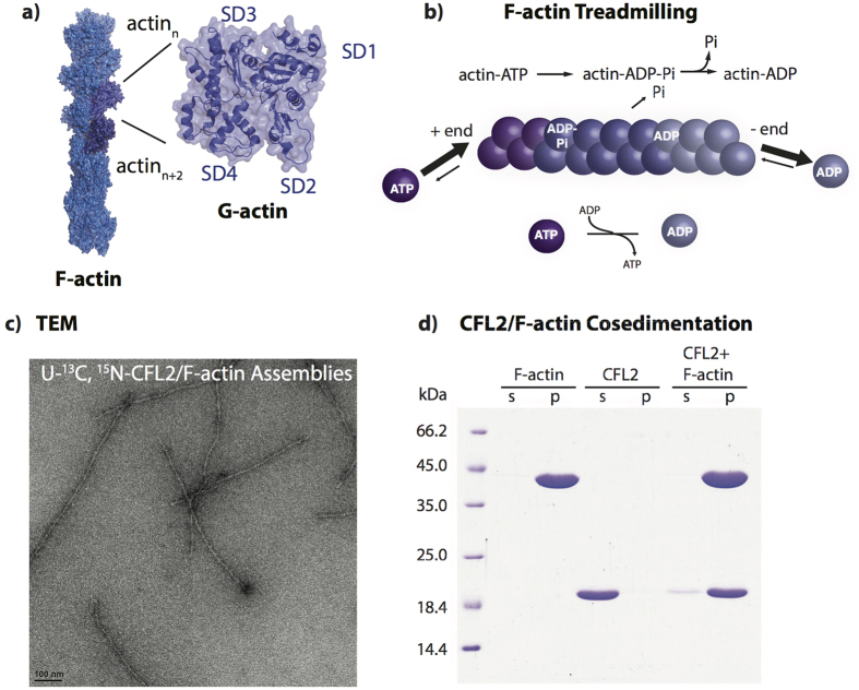
Figure 1.
(a) F-actin (blue) structure with G-actin protomers (“n” and “n + 2”) shown in purple. G-actin structure (purple) with subdomains labeled on the structure. (b) Actin treadmilling process for the polymerization and depolymerization of actin filaments. (c) TEM images of the NMR samples of U-13C,15N-CFL2 in complex with F-actin. (d) SDS-PAGE of CFL2/F-actin co-sedimentation. Samples containing either F-actin (42 kDa), CFL2 (18 kDa), or CFL2 complexed with F-actin were prepared under the conditions replicating the sample preparation for MAS NMR. Following ultracentrifugation, supernatants (s) and pellets (p) were resolved on SDS-gel.

