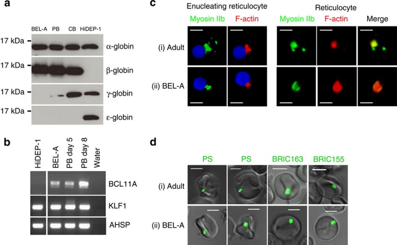
Figure 2. Characterization of BEL-A line.
(a) Western blots of late stage BEL-A, HiDEP-1 and erythroid cells differentiated from adult PB and cord blood CD34+ cells (at days 18, 12, 19 and 18 in culture, respectively) incubated with antibodies to α-, β-, γ- and ɛ-globin. (b) Expression of BCL11A and KLF1 transcripts in expanding BEL-A and HiDEP-1 cells and erythroid cells differentiated from adult PB CD34+ cells at days 5 and 8 in culture by RT-PCR. Transcripts for AHSP were included as a positive control. (c) Erythroid cells differentiated from adult peripheral blood CD34+ cells (i) and BEL-A cells (ii) were fixed, permeabilized and dual stained for F-actin (red) and myosin IIb (green). Single cells showing F-actin and myosin IIb localization in an enucleating cell, and in a reticulocyte are shown in three dimension. (d) Filtered adult reticulocytes (i) and BEL-A reticulocytes (ii) were live imaged after staining for phosphatidylserine (PS), glycophorin A intracellular domain (BRIC163) and band 3 intracellular domain (BRIC155) (all green). Scale bars 5 μm.

