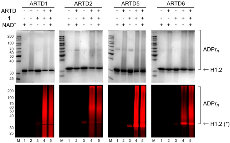
Figure 3.
SDS PAGE analysis of ADP-ribosylation of histone H1.2 with ARTD1, ARTD2, ARTD5 and ARTD6 using NAD+ analogue 1. Upper panel shows Coomassie Blue staining; lower panel shows TMR fluorescence. Experimental details are provided in Supporting Information File 1. *Unspecific staining of H1.2 in lanes 3 results from non-catalytic bond formation of NAD+ analogues with the protein.

