Figure 3.
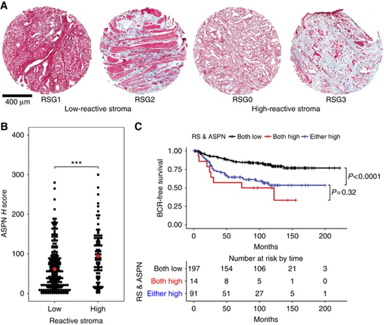
Co-identification of ASPN and reactive stromal histology. After IHC staining with ASPN and image storage, we stripped the TMA and performed Masson's trichrome staining to identify areas of reactive stroma. Low reactive stroma was scored as RSG1 and 2 while high reactive stroma was RSG0 and RSG3 (Ayala et al, 2003; Yanagisawa et al, 2007). (A) Representative images of samples graded as reactive stroma 1, 2, (low) and 0, and 3 (high) using Masson's trichrome. (B) Graph of patients showing ASPN staining and reactive stroma; ASPN staining was observed in patients with or without reactive stroma and there was evidence of elevated ASPN staining (H-score) and high reactive stroma (Low reactive stroma samples had a mean H-score of 61.5 while high reactive stroma samples showed an ASPN H-score of 94.3, t-test P=0.001). (C) Kaplan–Meier curve of progression among patients with no reactive stroma and low ASPN H-score (black), high reactive stroma and high ASPN (red), and patients showing either reactive stroma or ASPN (blue). Comparison among the three groups showed that samples positive for both reactive stroma and ASPN was predictive of progression as well as samples showing either high reactive stroma or ASPN (P<0.0001). There was no statistically significant difference between the double positive and single positive samples (P=0.32).

