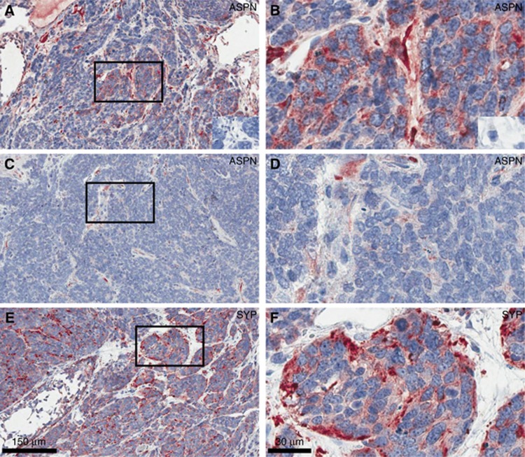
Figure 4.
Expression of ASPN in the murine TP53 prostate cancer model. Asporin was localised by immunohistochemistry in the mouse probasin driven TP53 prostate cancer model to determine whether there was expression in stromal cells and/or tumour cells. Prostate tumours from prostate specific p53 knockout mice showed ASPN staining in the stromal compartment (low magnification, A and C; high magnification, B and D). Tumours showed few stromal cells, though many of those present were positive for ASPN. IHC controls without primary antibody are insert in A and B. Left panels × 10 magnification and right panels at × 40 magnification. Synaptophysin staining is shown in E and F.

