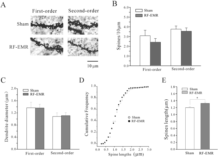
Figure 4. Effect of RF-EMR exposure on prefrontal cortical neuronal morphology observed by Golgi staining.
(A) Representative images of spine density on first- and second-order dendrites of cortical neurons in sham-exposed mice and at 3 days after 3.3 W/kg RF-EMR-exposure for 30 min. Scale bar is 10 μm. (B) Statistical analyses of spine density on first- or second-order dendrites of prefrontal cortical neurons in sham- and RF-EMR-exposed mice. (C) Effect of RF-EMR exposure on dendritic diameter. (D) Cumulative frequency distributions of spine length in sham- and RF-EMR-exposed mice were compared using Kolmogorov–Smirnov tests. (E) Statistical analyses of spine length on first- and second-order dendrites of mPFC neurons in sham- and RF-EMR-exposed mice. *p < 0.05 for two groups connected by a straight line using a two-sample t-test.

