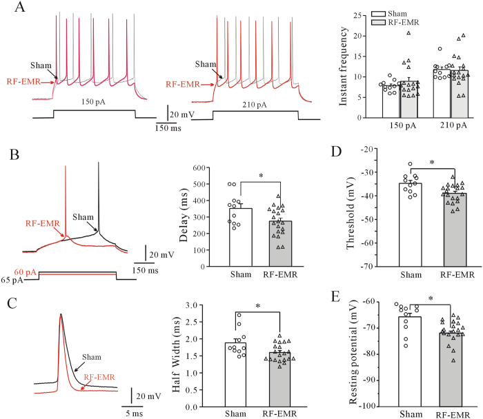
Figure 5. Effect of RF-EMR exposure on action potentials (APs) and membrane potentials in prefrontal cortical neurons.
(A) Representative current-clamp recording and statistical analyses of AP frequency of cortical neurons in sham- and RF-EMR-exposed mice. (B,C) Representative sample and statistical analyses showing effect of RF-EMR exposure on delay of first AP elicited by current injection, and half-width of AP in sham- and RF-EMR-exposed mice. (D,E) Effect of RF-EMR exposure on resting membrane potential and threshold potential in cortical neurons in sham- and RF-EMR-exposed mice. *p < 0.05, for two groups connected by a straight line using a two-sample t-test.

