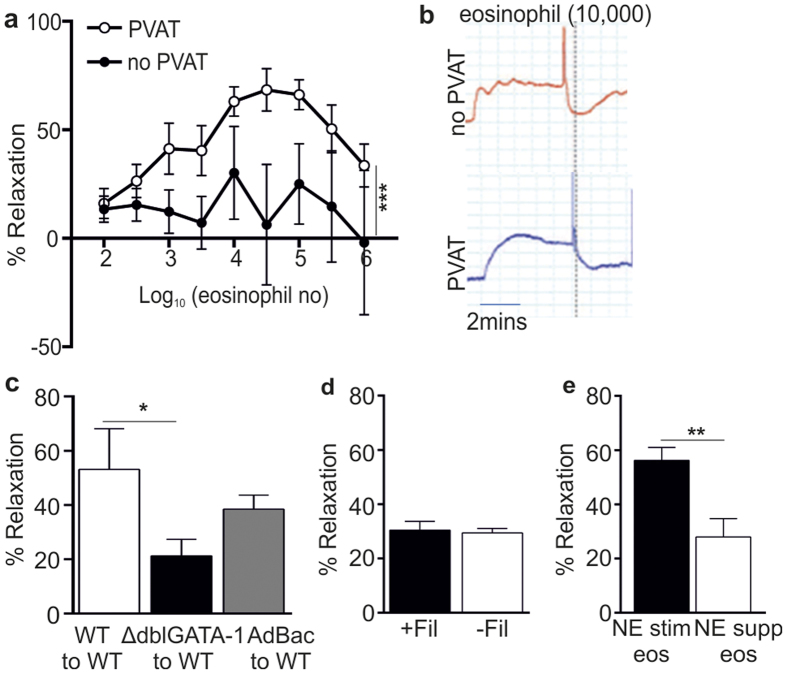
Figure 3. Exogenous application of purified eosinophils induces vessel relaxation.
Isolated splenic eosinophils were added to mesenteric arteries preconstricted with norepinephrine (10−5 M). Relaxation was measured by wire myography. (a) Exogenous application of eosinophils induced dose-dependent relaxation of ΔdblGATA-1 arteries in the presence (-ο-) but not absence (-•-) of PVAT (n = 8, pooled from 8 individual experiments; ***P < 0.0001, two-way ANOVA). (b) Trace representative of a pre-constricted ΔdblGATA-1 artery ± PVAT responding to 10,000 eosinophils. Note the rapid drop in constriction following addition of eosinophils (dotted line). (c) Solution transfer experiments to assess generation of a transferable factor that mediates relaxation (n = 10, pooled from 2 experiments; *P = 0.049, Student’s t-test. (d) Vessel relaxation in response to application of NE-stimulated eosinophils (−Fil) or filtered supernatant from NE-stimulated eosinophils (+Fil), by passing through a 0.2 μm filter to retain eosinophils, to pre-constricted WT mesenteric arteries in the presence of PVAT. (e) Addition of NE-stimulated (10−5 M) eosinophil supernatant or unstimulated eosinophil supernatant supplemented with NE (10−5 M) to pre-constricted ΔdblGATA-1 arteries in the presence of PVAT (n = 6; **P = 0.0068, student’s t-test).

