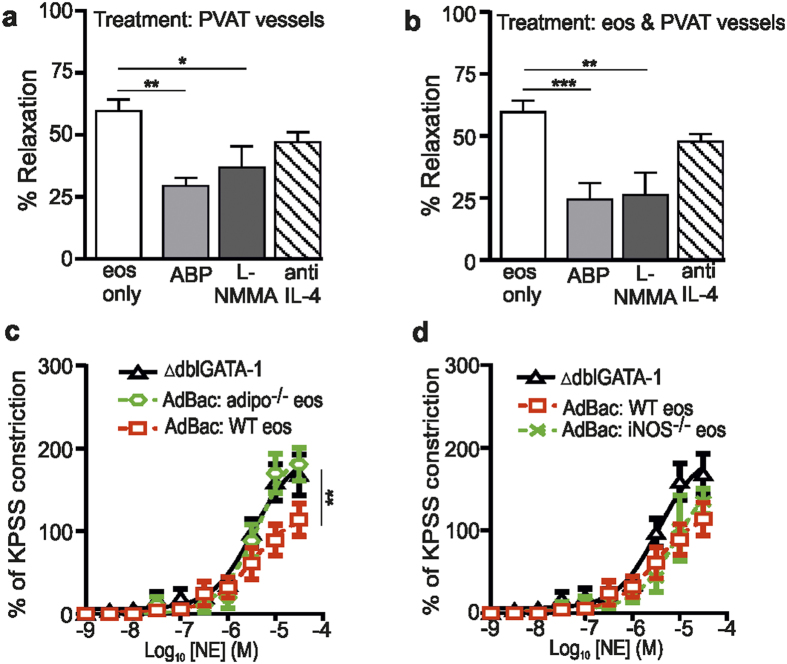
Figure 4. Eosinophils induce adiponectin- and NO-dependent PVAT-induced vessel relaxation.
The functional role of adiponectin and NO were investigated by wire myograph analysis of mesenteric vessels of ΔdblGATA-1 mice using pharmacological tools and in eosinophil-reconstituted ΔdblGATA-1 mice. (a,b) Vessel relaxation in response to exogenous application of 10,000 NE-stimulated eosinophils (eos) to preconstricted ΔdblGATA-1 mesenteric arteries + PVAT following incubation of (a) PVAT (L-NMMA, anti-IL-4: n = 9, ABP: n = 8; *P < 0.05 and **P < 0.01, one-way ANOVA with post hoc Dunnett’s), or (b) PVAT and eosinophils (L-NMMA, ABP, anti-IL-4: n = 9; **P < 0.01, ***P < 0.001, one-way ANOVA with post hoc Dunnett’s). (c,d) The PVAT anti-contractile effect of NE constricted arteries from ΔdblGATA-1 mice reconstituted with (c) adiponectin−/− (- -), (c,d) WT (-□-), or (d) iNOS−/− (-×-) eosinophils (adipo−/− vs WT: n = 4, **P = 0.0085, two-way ANOVA; iNOS−/− vs WT: n = 5; P = NS, two-way ANOVA).

