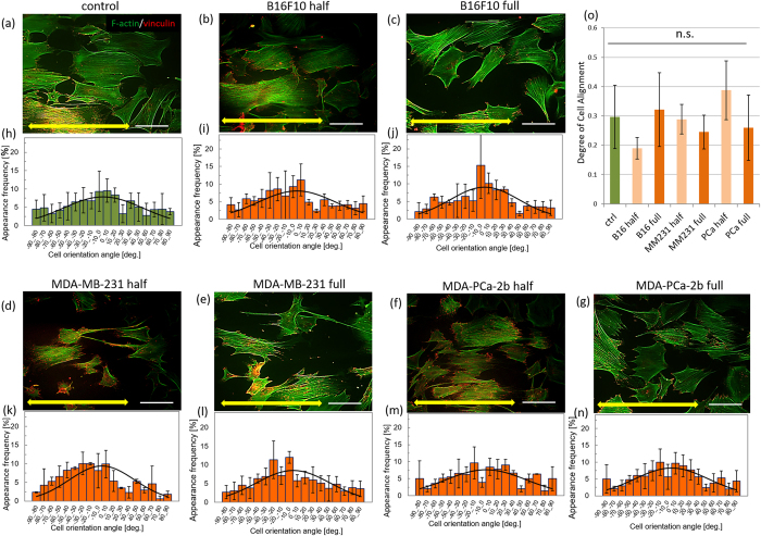
Figure 3.
Immunocytochemical analysis of osteoblasts cultured with cancer cell-conditioned media ((a): control, (b,d,f): 50% concentration, and (c,e,g): 100% concentration of (b,c): mouse melanoma B16F10, (d,e): human breast cancer MDA-MB-231, (f,g): human prostate cancer MDA-PCa-2b cells conditioned media). The histograms of cell angular distribution are shown in figures (h–n). The osteoblasts are preferentially aligned along the substrate collagen orientation (yellow arrows). (o) The degree of cell alignment along the substrate collagen orientation. Osteoblast alignment is not significantly affected by the cancer cell-derived conditioned media. Scale bar: 100 μm.

