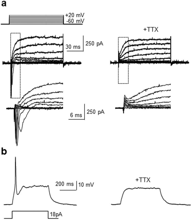
Figure 4. Sin3A-silenced P19 cells are differentiated into electrophysiologically active neurons.
Sin3A-knockdown P19 cells were incubated for 21 days. (a) Whole-cell patch clamp recording of differentiated cells reveals generation of fast activating currents induced by 10 mV depolarizing voltage steps from −60 mV to + 20 mV before (left) and after (right) 0.5 μM tetrodotoxin (TTX) perfusion (n = 4/7 of recorded cells). Insets show respective traces on an expanded scale. (b) Action potentials generated by current injection in the differentiated cells (left) before and (right) after 0.5 μM TTX perfusion (n = 4/7 of recorded cells).

