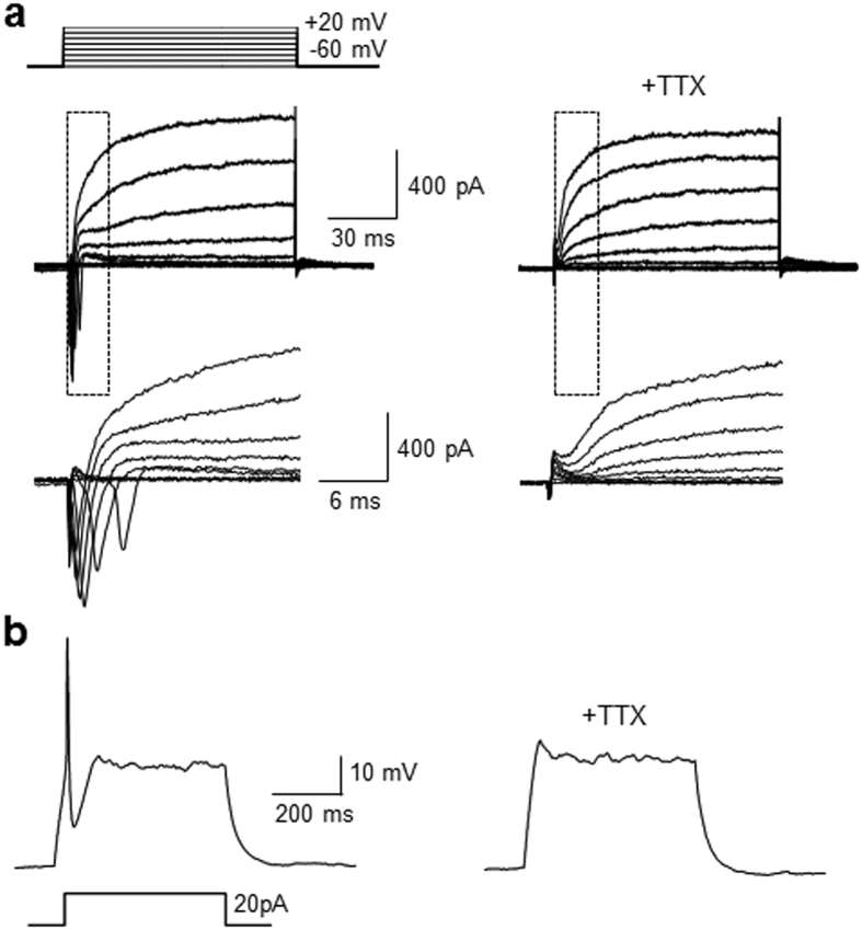
Figure 7. Neurogenic cells differentiated from P19 cells after treatment with TAT-NLS-Mad1 exhibit electrophysiological properties.
P19 cells were incubated with 30 μM TAT-NLS-Mad1 for 21 days. (a) Representative traces of whole-cell current-clamp induced by 10 mV depolarizing voltage steps from −60 mV to +20 mV before (left) and after (right) 0.5 μM tetrodotoxin (TTX) perfusion (n = 5/12 of recorded cells). Insets show respective traces on an expanded scale. (b) Representative traces of an action potential recorded in differentiated Sin3A-knockdown cells in the current-clamp mode in response to depolarization by current injection before (left) and after (right) 0.5 μM TTX perfusion (n = 5/12 of recorded cells).

