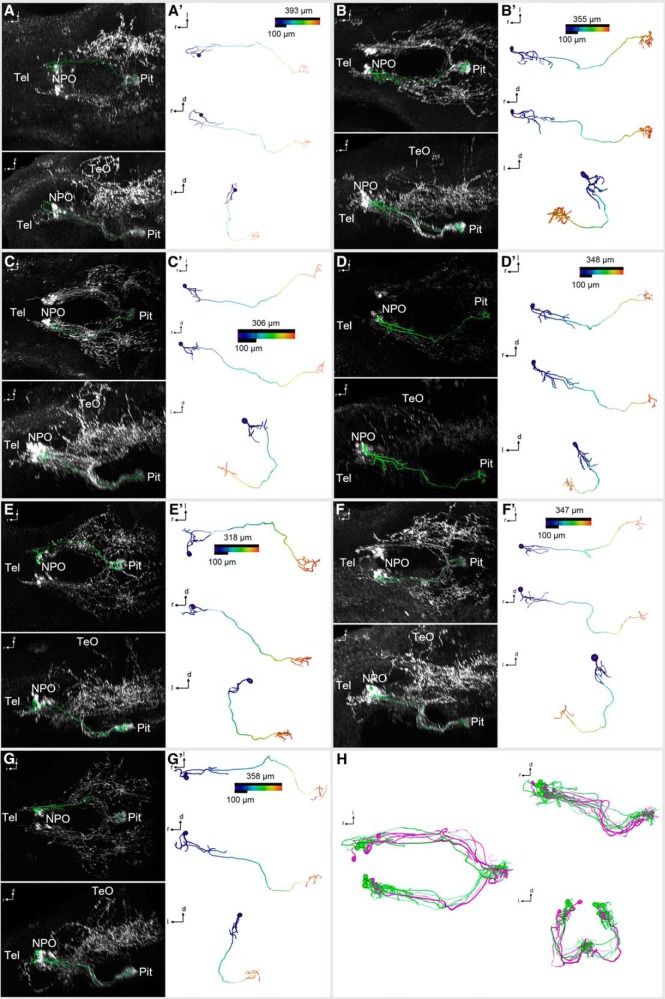
Figure 6.
3D reconstruction of morphologically complex Oxt-positive preoptic neurons that innervate the pituitary at 6 dpf. A–G″, Skeletonized neurons, shown within the context of the Oxt IHC (A–G; dorsal and lateral views), and shown individually with a color code of the distance along each branch from the soma center (A′–G′; dorsal, lateral, and frontal views). Distance color codes are scaled to the maximum distance of each cell. Note that these cells feature several branches close to the soma. H, Registration of the hypophysiotropic cells shown in Fig. 5 (magenta) and Fig. 6 (green) shows intermingled projections (dorsal, lateral, and frontal views).

