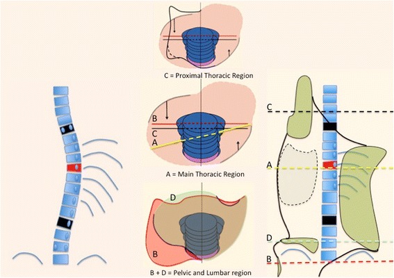
Fig. 6.

This figure shows the corrective principles for a single long-low thoracic curve with the apical vertebra still in the main-low thoracic region (described later as A1 type in Rigo classification): “regional derotation” and “three-point system.” The region of the trunk affected by the single structural curve is over-derotated to the left (yellow line A) throughout a dorsal-lateral pad and a ventral pad, against the two caudal and cranial regions. Pelvis and lower lumbar regions (B + D) are fixed in the frontal plane of reference (0° of rotation). The pelvis section of the brace is asymmetric, with the lateral-dorsal part opened in the right side and supported by left lumbar contact as well as anterior abdominal contact. The proximal thoracic region (C) is also fixed in the frontal plane of reference with a dorsal left counter-rotation pad. A left lateral to medial pad acts in the proximal thoracic region as the third proximal point of the “three-point system.” The lateral component of the dorsal-lateral pad is the second point, on the right side. The left pelvis section together with the lateral component of the left lumbar support acts as the first caudal point of the system. The brace provides a left lateral-dorsal and a ventral right expansion rooms to facilitate breathing expansion and growth. The dorsal-lateral and anterior pads forming the pair of forces for derotation work both at the same level (maximum force at the apical level). This original design—A1 type—has shown to produce the highest percentage of in-brace correction [64]
