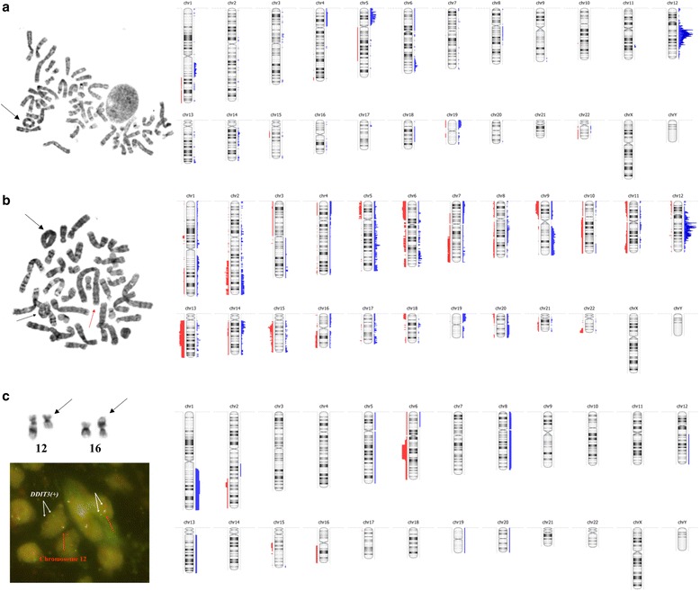
Fig. 3.

Comparison of the standard diagnostic approach and array-CGH in WDLPS, DDLPS and MLPS tumors. On the left the conventional karyotyping and/or FISH results are shown. Black arrows depict the supernumerary ring chromosomes in WDLPS and DDLPS tumors (a–b) and the balanced translocation t(12;16) in MLPS sample (c). DDIT3 break apart probe was used in FISH analysis (one orange and one green signal pattern indicate a rearrangement of the DDIT3 gene region). On the right the overview of all copy number aberrations (CNAs) detected by array-CGH is presented. Red and blue colors represent losses and gains, respectively
