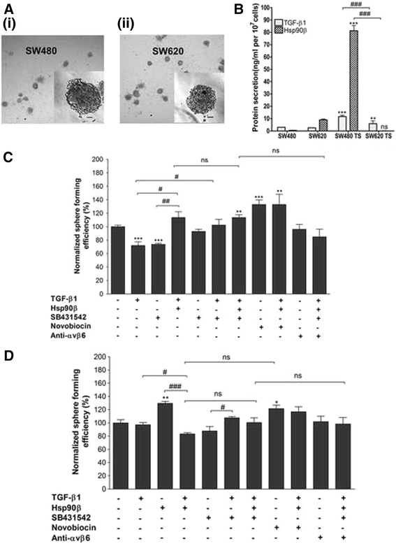
Fig. 5.

Investigation of the role of TGF-β and Hsp90 in anchorage-independent growth of SW480 and SW620 cells. a Photograph of tumourspheres formed by SW480 (i) and SW620 (ii) cells taken under a light microscope at 100x magnification. Scale bars indicate 100 μm. b Comparison of TGF-β1 and Hsp90β secretion by SW480 primary and SW620 secondary tumour-derived cells grown adherently and in suspension using a DuoSet ELISA kit (R and D systems) and sandwich ELISA method, respectively. Data shown are representative of three individual experiments carried out in triplicate and showing consistent results. Statistical significance was assessed using GraphPad Prism 4.03 software by means of a two-way analysis of variance (ANOVA) with Bonferroni post-test where n = 3. Comparisons in terms of the levels of proteins between adherent cells and tumoursphere (within cell lines) is indicated by asterisks (*), while comparisons between cell lines is indicated by hashes (#). c and d Analysis of the effect of addition or inhibition of TGF-β or Hsp90 on tumoursphere formation. Sphere forming efficiency (percentage of the total number of cells seeded that are able to form tumourspheres after 7 days) of SW480 c and SW620 cells d was normalized to that of untreated cells for each cell line (taken as 100%). Error bars indicate the standard error in the mean where n = 4. Statistical significance was assessed using GraphPad Prism 4.03 software by means of a two-way analysis of variance (ANOVA) with Bonferroni post-test and significance between untreated cells and those after each treatment (indicated by asterisks) as well as between particular treatments (indicated by hashes) are shown (*p < 0.05, **p < 0.01, # p < 0.05, ## p < 0.01, ### p < 0.001)
