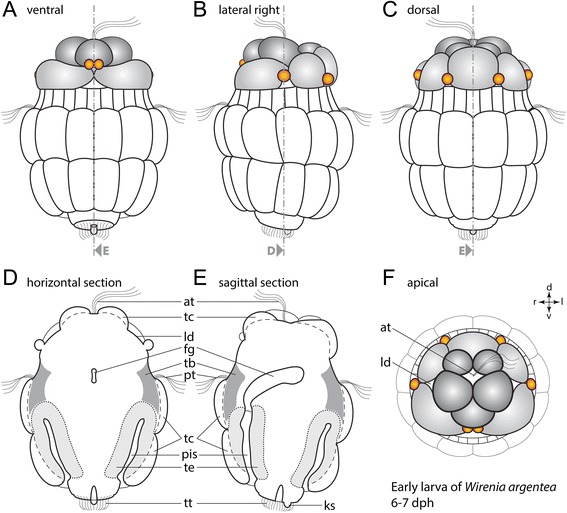
Fig. 3.

Line drawings of gross morphology of the early W. argentea larva. Schematic drawing of an early test cell larva (6-7 days post hatching) based on autofluorescence confocal scans. Anterior faces up in (a)-(e) and towards the viewer in (f). a Ventral view. b Lateral view from the right. c Dorsal view. d Horizontal section. e Sagittal section. f Anterior view of the episphere showing lateral depressions. Note the characteristic cell arrangement of two rows of test cells in the hyposphere, two rows of test cells with opposing configuration in the episphere (grey/dark grey), and six bilaterally arranged cells or cell protuberances (orange) in the episphere. Dorsal (d)–ventral (v) and left (l)–right (r) axes indicate the orientation. Abbreviations: apical tuft (at); lumen of foregut (fg); posterior knob-like structure (ks); lateral depression (ld); peri-imaginal space (pis); prototroch (pt); trochoblast (tb); test cells (tc); epidermis of the trunk (te); telotroch (tt)
