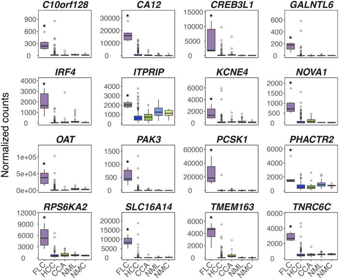
Figure 3. Sixteen genes most uniquely distinguish FLC from other liver cancers.
Boxplots depicting RNA levels of 16 genes in the FLC mRNA signature in FLC (n = 6), HCC (n = 263), CCA (n = 36), non-malignant liver (NML, n = 50), and non-malignant cholangiocyte/bile duct (NMC, n = 9) from TCGA. Y-axis shows counts normalized by DESeq. Shaded regions of boxplots show the 25th–75th quantiles of the data with the median denoted by a bold line. Whiskers of boxplots represent data <25th and >75th quantiles. Circles represent data points that are outliers, defined as points <25th quantile minus 1.5*IQR (interquartile range, 75th–25th quantile) or >75th quantile plus 1.5*IQR. *FDR < 0.05 (DESeq, negative binomial test) of FLC compared to both HCC and CCA.

