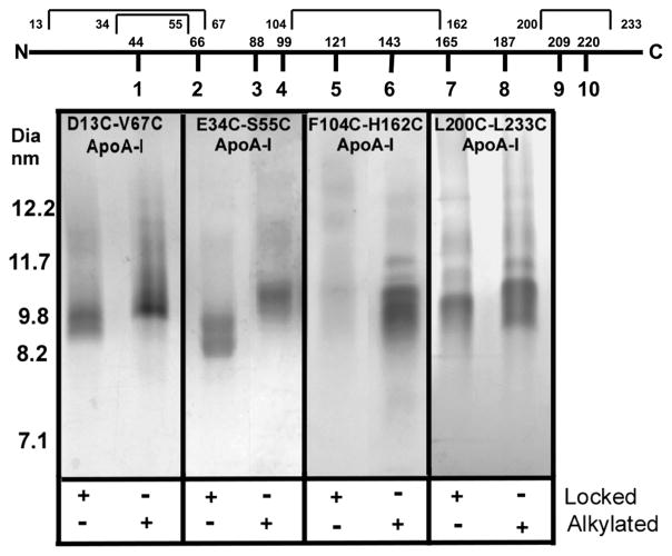
Figure 2.
Coomassie Blue-stained 4 to 30% NDGGE of rHDL particles formed from cholate dialysis of (80:1 molar ratio) POPC and apoA-I. Locked indicates the presence (+) or absence (−) of a disulfide bond. Alkylated indicates the presence (+) or absence (−) of carbamidomethylated cysteines. The top part of the figure shows apoA-I divided into its 10 repeats with the first amino acid in the repeat displayed. The intramolecular disulfide bonds are indicated above with the position of the first and last cysteine.

