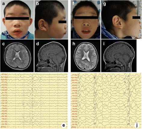
Fig. 2.

Clinical features of patient 1 and 2. Clinical features of patient 1 (a-e). Facial dysmorphisms with brachycephaly, slightly upturned nares, large and low set ears were observed (a, b). Axial T2 (c) and sagital T1 (d)-weighted brain MRI images were in normal limits. interictal EEG shows epileptic sharp wave discharges from the right temporal cerebral area with poorly regulated posterior rhythm and slow background activity (e). Clinical features of patient 2 (f-j). Facial dysmorphisms with large ears, slightly upturned nares, and midface hypoplasia followed by depressed nasal bridge were observed (f, g). Axial T2 (h) and sagital T1 (i)-weighted brain MRI images showed no abnormalities. interictal-EEG reveals frequent generalized burst of epileptic sharp wave discharges from the both frontal cerebral area followed by attenuation of background activity (j)
