


Figure 2. WD does not affect Aβ loads in brain parenchyma.
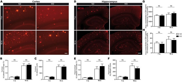
Fluorescent 6E10 immunostaining for Aβ plaques in the A. cortex or D. hippocampus of APPswe/PS1 mice. Quantification by stereological analyses of B., E. size (μm2) and C., F. number of 6E10-positive Aβ plaques in the A.-C. cortex and D.-F. hippocampus of 7 and 16 months old ND or WD-fed animals. No significant changes were observed neither in the cortex nor the hippocampus of WD-fed animals in comparison to ND-fed littermates, in terms of Aβ plaques B., E. size and C., F. number. Soluble Aβ species were measured by ELISA in brain (TBH) G., H.. No significant changes were observed in soluble G. Aβ 1-42 or H. Aβ 1-40 levels normalized per mg of total proteins. Data are means ± SEM (n = 10-12 animals per group). Cortical Aβ plaques size; Two-way ANOVA p = 0.4309 and cortical Aβ plaques number; Two-way ANOVA p = 0.9430, Bonferroni post-hoc tests p > 0.05. Hippocampal Aβ plaques size; Two-way ANOVA p = 0.2531 and hippocampal Aβ plaques number; Two-way ANOVA p = 0.2115, Bonferroni post-hoc tests p > 0.05. Soluble Aβ 1-42; Two-way ANOVA p = 0.7177 and Aβ 1-40; Two-way ANOVA p = 0.6373, Bonferroni post-hoc tests p > 0.05. Scale bar A = 100μm. Scale bar D = 250μm.