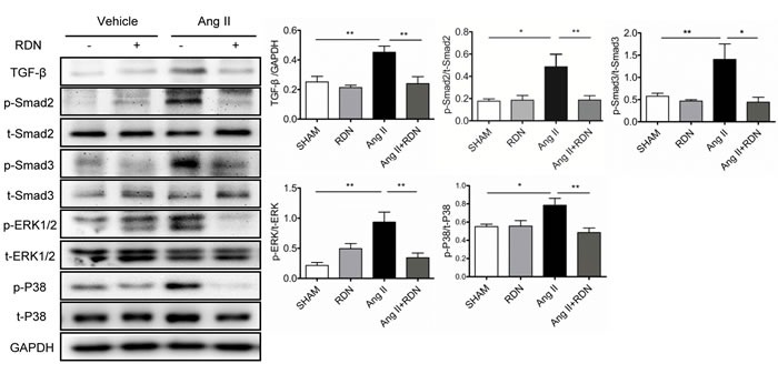
Figure 6. Suppression of TGF-β-Smad2/3 and MAPKs in RDN rats with Ang II stimulation.

Cardiac protein expression of TGF-β, Smad2 phosphorylation, Smad3 phosphorylation, ERK1/2 phosphorylation and p38 phosphorylation. Anti-GAPDH antibody served as a loading control. Values are mean ± SEM. n = 4-8 in each group. *P < 0.05, **P < 0.01.
