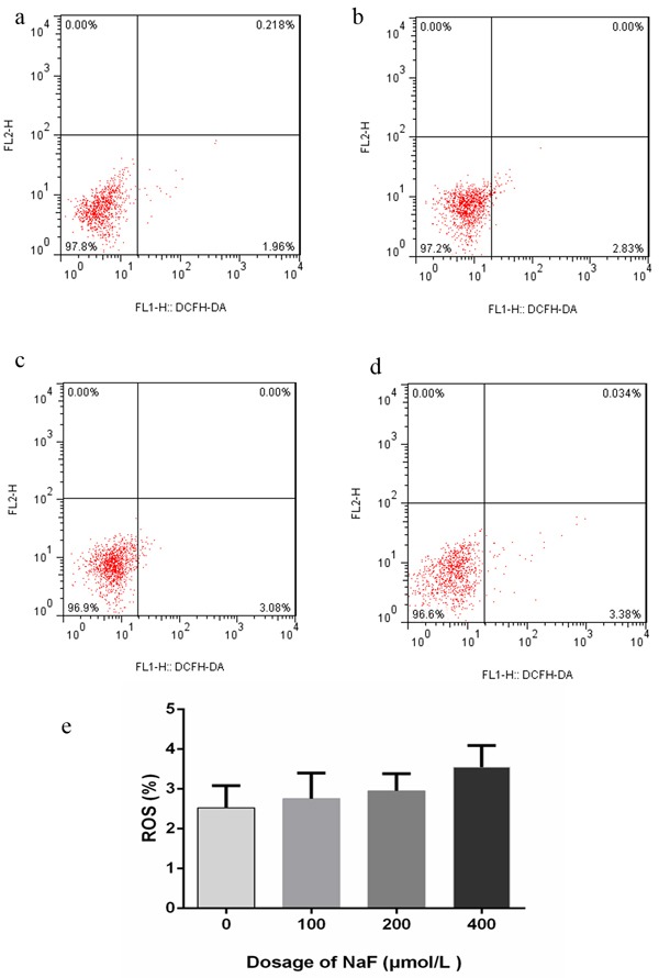
Figure 2. Effect of NaF on ROS generation at 24 h.

a.-d. Two-dimension scatter plots depicting distribution of cells positively stained for DCFH-DA. (a) CG, (b) LG, (c) MG and (d) HG. e. Quantitative analysis of ROS generation. Data are presented with the means ± standard deviation, * p < 0.05, ** p < 0.01, compared with the control group. Data were analyzed by the variance (ANOVA) test of the SPSS 19.0 software.
